Monday, June 29, 2020

Plus Two Biology|Zoology|Chapter-6|Molecular Basis of Inheritance|Part-VI|Genetic code|Translation

#biostripsmedia# #pratheeshpallath#

In this session Bio Strips Media takes through the important topics of Molecular Basis of Inheritance. It is one of the most important and scoring topics in Plus Two Biology Exam and NEET Exam. 


This lecture covers:

·         Genetic code

·         Translation


GENETIC CODE

Genetic code refers to the relationship between the sequence of nucleotides in mRNA and the sequence of amino acids in the polypeptide.

George Gamow, who suggested that the code must be made up of three bases(triplet codons).

The 64 distinct triplets determine the 20 amino acids. These triplets are called codons. Any coded message by these codons is commonly called cryptogram.

Har Gobind Khorana could synthesise RNA molecules with definite combinations of bases.

Marshal Nierenberg made a cell free system for protein synthesis that helped in deciphering the code.

Severo Ochoa discovered enzyme polynucleotide phosphorylase that could polymerise RNA with definite sequence in template - independent manner.   

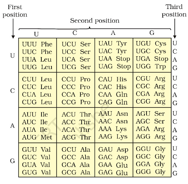
The checkerboard pattern of genetic code was prepared as given below:


The salient features of genetic code are given below:

The codons are triplets and there are 64 codons, 61 codons code for 20 amino acids and 3 codons do not code for any amino acid, but function as stop/termination codons or nonsense codons. 

Three codons viz UAG (Amber), UAA (ochre) and UGA (opal) are non sense codons.

Each codons code for only one amino acid and so the genetic code is unambiguous and specific.

Since some amino acids are coded by more than one codon, the genetic code is said to be degenerate.

The codons are read in a contiguous manner in a 5' -3' direction and have no punctuations.

The genetic code is universal, the codons code for the same amino acid in any organism.

AUG has dual functions of coding for methionine and acting as initiation codon. 

Wobble hypothesis was given by FHC Crick . According to this,  the third nitrogenous base of a codon is not much significant and Codon is specified by the first two bases. Hence the same tRNA can recognise more than one codons differing only at third position. 

Translation

It is a process in living cells in which the genetic information encoded in messenger RNA in the form of a sequence of nucleotide triplets (codons) is translated into a  sequence of amino acids in a polypeptide chain during protein synthesis.

Ribosomes serve as the site for protein synthesis hence they are called protein factories. Each ribosome consists of large and small subunits.  the two subunits come together only at the time of protein synthesis. The phenomena is called Association . MG 2 + is essential for it. Many ribosomes line up on the mRNA chain during Protein synthesis. Such a row of active ribosomes is called a polyribosome or simply a polysome.

Ribosome also acts as a catalyst (23 s r RNA in bacteria is enzyme ribozyme) for formation of peptide Bond.

Eukaryotic ribosome has a groove at the junction of the two subunits. Trom this groove a tunnel extends through the large subunit and opens into a Canal of  the endoplasmic reticulum.  The polypeptides are synthesized in the groove between the two ribosomal subunits and pass through the Tunnel of the large subunit into the endoplasmic reticulum. While in the groove the developing polypeptide is protected from the cellular enzymes. The small subunit forms a cap over the large subunit. The large subunit attaches to the endoplasmic reticulum by two glycoproteins named ribophorin 1 and 2.

A ribosome has one binding site for mRNAi and three binding sites for t RNA. P- site (peptidyl tRNA site) that holds the tRNA carrying the growing polypeptide chain. A- site (aminoacyl tRNA site) which holds the tRNA carrying next  amino acid to be added to the chain and E -site (exit site) were discharged tRNAs leave the ribosome. these sites span across the large subunits of the ribosome. however the first tRNA amino acid Complex directly enters the P site of the ribosome.

It is a messenger RNA which brings coded information from DNA and takes part in its translation by bringing amino acids in a particular sequence during the synthesis of polypeptide. the codons of mRNA are recognised by anticodons of tRNAs. 

Protein synthesis is an elaborate process and occurs as follows

Activation of amino acids

In this process amino acid reacts with ATP to form amino acid amp Complex and pyrophosphate. This complex is called an activated amino acid.

The activated amino acid attached to the amino acid binding site (3'end) of its specific t RNA. where its cooh group bonds to 0h group of the terminal base triplet CCA. The process commonly called as charging of tRNA or aminoacylation of tRNA.

The reaction is catalysed by the enzyme amino acyl tRNA synthetase.

Initiation of polypeptide chain

The translation of mRNA begins with the formation of initiation complex. The union of mRNA,a tRNA bearing the first amino acid of the polypeptide and the two subunits of a ribosome forms the translation initiation complex.

Proteins called initiation factors IFs in prokaryotes and eIFs in eukaryotes are necessary to bring all the components of the translation initiation Complex together. GTP supplies the energy for the formation of initiation complex.

The small subunit of a ribosome attaches to mRNA and specific charged initiator tRNA. 

In prokaryotes rRNA of the small subunit base pairs with specific sequence of nucleotides in the mRNAs leader segment.

 in eukaryotes 5Prime cap first tell the small subunit to bind to the mRNAs five Prime end.

The initiator codon (AUG) which signals the start of translation

The initiator tRNA joins the initiation codon by its anticodon through hydrogen bonds.

It carries  the amino acid formyl methionine in prokaryotes and methionine  in eukaryotes.

Now the large subunit of ribosome joins the small subunit.

At this stage the initiator tRNA lies at the P site of the ribosome and the A site is vacant to let another charged tRNA to enter.

Elongation of polypeptide chain

In the elongation stage of translation, amino acids are added one by one to the first amino acid. Each addition requires the help of certain proteins called elongation factors. In prokaryotes the elongation factors are termed EF-Tu, EF-Ts and EF-G.

An aminoacyl tRNA with its amino acid enters the ribosome at the A site. It's anticodon reads and binds to the complementary codon of mRNA by hydrogen bonds.

The amino acid on tRNA at P site and the newly arrived amino acid at A site join by a peptide Bond. It is formed between the carboxyl group of first amino acid and the amino group of the second amino acid. the reaction is catalysed by peptidyl transferase.

Thus the second tRNA carries a dipeptide and the first t RNA is without amino acid.

Translocation -  in this step the ribosome moves one codon towards the three Prime end of the mRNA. 

This movement shifts the anti codon of the dipeptidyl tRNA from the A site to the P site and shifts the deacylated tRNA from the P site to the E site, from where the deacylated tRNA is released into the cytosol.

The third codon of the mRNA now lies in the A site and the second codon in the P site.

Now the next cycle starts. The ribosome moves from codon to codon along the mRNA in the five Prime to 3 Prime direction.

 Amino acids are added one by one in the sequence of the codon and become joined together to form polypeptide.

Termination of polypeptide synthesis

Termination is signaled by the presence of one of three termination codons in the mRNA (UAA, AUG, UGA) imediately following the final coded amino acid. 

When one of the  termination codon comes at A site it does not code for any amino acid and there is no tRNA molecule for it. as a result the polypeptide synthesis stops.

 Three  release factors RF-1, RF-2 and RF-3 contribute in release of the free polypeptide and last tRNA from the ribosome. 

The process is repeated several times creating polysomes. mRNA has some additional sequences that are not translated are referred to as untranslated regions (UTRs), required for efficient translation.


Wednesday, June 17, 2020

MOLECULAR BASIS OF INHERITANCE|PART-V|TRANSCRIPTION


#biostripsmedia# #pratheeshpallath#

In this session Bio Strips Media takes through the important topics of Molecular Basis of Inheritance. It is one of the most important and scoring topics in Plus Two Biology Exam and NEET Exam. 


This lecture covers:

·         Central dogma of molecular biology

·         Transcription in prokaryotes

·         Transcription in eukaryotes

·         mRNA processing


The content being discussed in this video will be helpful for those appearing for the Plus Two Biology Exam and NEET Biology Exam.


The information in this video is very useful to them and it helps those NEET aspirants to score maximum marks in Biology.


Central dogma of Molecular  biology

Crick (1958) Proposed the central dogma of molecular biology. Central dogma is the unidirectional flow of information from DNA to RNA and from RNA to polypeptide. H. Remind and Baltimore (1970) Introduced the concept of reverse central Dogma, ie, formation of DNA from RNA. it is also called Teminism and of occurs in retroviruses. Many tumour viruses contain RNA as genetic material and replicate by first synthesizing a complimentary DNA. this process  is called reverse transcription. It is carried out by an RNA dependant DNA polymerase called reverse transcriptase.             

Transcription

The process of transferring information stored in DNA to RNA through the synthesis of mRNA over the template of DNA is called transcription. Transcription Occurs in the nucleus during G1 and G2 phases of cell cycle. A transcription unit in DNA has three regions:

·        A promoter

·       Structural gene and

·       A terminator

The process is catalysed by DNA-dependent RNA-polymerase, which catalyses the polymerization of nucleotides only in 5' - 3' direction. 

The DNA strand with 3' - 5' polarity is called template strand while the other strand with 5' - 3' polarity is called coding strand.

The promoter refers to a particular sequence of DNA located towards the 5'end of the coding strand, where the RNA polymerase becomes bound for transcription.

The terminator is a sequence of DNA located towards the 3' end of the coding strand, where the process of transcription stops. 

Only one of the strands of the DNA act as the template for RNA synthesis for the following reasons:

If the both strands code for RNA, two different RNA molecules and two different proteins would be formed. 

Since the two RNA molecules produced would be complementary to each other, they would wind together to form a double stranded RNA without carrying out translation.

Transcription in prokaryotes

In bacteria transcription takes place in three steps which are initiation, elongation and termination.

In prokaryotes the structural genes are polycistronic and continuous.

In prokaryotes there is a single DNA-dependent RNA-polymerase that catalyses the transcription of all the three types of RNA (mRNA, tRNA, rRNA).

RNA polymerase binds to the promoter and initiates the process along with certain initiation factors (sigma factor). The sigma factor Recognises the promoter region while the remaining core enzymes take part in the transcription.

The initiation also facilitates opening of the helix and continues elongation.

It uses ribonucleoside triphosphate for polymerization on a DNA template following complementarity of bases.

Once the RNA polymerase reaches the terminator the RNA polymerase falls off and the nascent RNA separates; it is called termination of transcription and is facilitated by certain termination factors (rho factor).

In prokaryotes the mRNA synthesized does not require any processing to become active and both transcription and translation occur in the same cytosol; translation can start much before the mRNA is fully transcribed, ie, transcription and translation can be coupled.  

TRANSCRIPTION IN EUKARYOTES

In eukaryotes the structural genes are monocistronic and split.

They have coding sequence called exons that form part of mRNA and non-coding sequences called introns, that do not form part of the mRNA and are removed during splicing.

In eukaryotes there are at least three different RNA polymerases in the nucleus, which function as follows:

·       RNA polymerase I transcribes rRNAs

·       RNA polymerase II transcribes mRNAs (hnRNA) and small nuclear RNAs.

·       RNA polymerase III transcribes tRNAs and small ribosomal RNAs.

The primary transcript contains both exons and introns and it is subjected to a process called splicing, where the introns are removed and exons are joined in a definite order to form mRNA.

The hnRNA undergoes two additional processes called capping and tailing.

In capping methyl guanosine tri-phosphate is added to the 5' end of hnRNA.

In tailing adenylate residues (about 200-300) are added at the 3' end of hnRNA.

The fully processed hnRNA is called mRNA and is released from the nucleus into the cytoplasm.

This processing helps in keeping mRNA away from the degrading action of nucleases present in nucleus.


Tuesday, June 16, 2020

KERALA PSC BASIC SCIENCE PREVIOUS QUESTIONS -PART-VI


#biostripsmedia# #pratheeshpallath#

The Important questions asked by Kerala PSC from Basic Science are mainly discussed in this video.

The SCERT Text Book Based facts are very useful for all competitive exams like LP-UP/LDC/LGS/SI/BDO/Police Constable etc.

In this video I explain all those important facts related to the Previous questions. This video is useful to score maximum marks for various competitive exams. 

Previous Questions|Kerala PSC|Basic facts|Basic Science|Biology|Physics|Chemistry|Kerala PSC Coaching Class Malayalam|K TET Coaching Class|EVS|Category I|CategoryII

The content being discussed in this video will be helpful for those appearing for the K-TET Exam. EVS is the integral part of K-TET Category I and II Exams. The information in this video is very useful to them and it helps those K-TET aspirants to score maximum marks in EVS.

The relevant facts discussed in this video course is useful to prepare for LP/UP Assistant Exam 2020 and LDC 2020.

The Science content in this video will be helpful for those candidates appearing for various Lower Division Clerk Exams, University Assistant Exam, Last Grade Servants Exam, Company Corporation Board Assistant Exams, Secretariat Assistant Exams, Sub Inspector Exam and all other Exams conducted by Kerala Public Service Commission (Kerala PSC) and various other agencies. 

To get more videos from Bio Strips Media related to Kerala PSC Coaching and K-TET Coaching do subscribe to our channel and receive notification by clicking the Bell icon.

For more videos and updates please visit our channel.

Follow me On:


https://www.facebook.com/biostripsmedia

 https://www.instagram.com/bio_strips_media/

https://biostripsmedia.blogspot.com


Wednesday, June 10, 2020

MOLECULAR BASIS OF INHERITANCE - PART-IV|REPLICATION


#biostripsmedia# #pratheeshpallath#

In this session Bio Strips Media takes through the important topics of Molecular Basis of Inheritance. It is one of the most important and scoring topics in Plus Two Biology Exam and NEET Exam. 


This lecture covers:

·         Replication of DNA

·         Enzymes involved in DNA replication

·         Mechanism of DNA replication


The content being discussed in this video will be helpful for those appearing for the Plus Two Biology Exam and NEET Biology Exam.


The information in this video is very useful to them and it helps those NEET aspirants to score maximum marks in Biology.


The Biology content in this video will be helpful for those candidates appearing for Kerala SET Zoology Exam, Kerala SET Botany Exam, KTET Natural Science Exam and various other Entrance Exams. 


DNA Replication

Replication is the process of formation of carbon copies or duplication of DNA.

Replication in eukaryotes occurs in the nucleus during the 'S' phase of the cell cycle. In prokaryotes replication takes place in the cytoplasm. Watson and Crick suggested that the two strands of DNA molecule uncoil and separate and each strand serves as a template for the synthesis of a new (complementary) strand.

Two daughter DNA molecules are formed from the parent DNA molecule and  these are identical to the parent molecule. Each daughter DNA molecule consists of one parent strand and one new strand. Since only one parent strand is conserved in each daughter molecule, this mode of replication is said to be e semi-conservative. Semiconservative replication of DNA a was found by Taylor (1957) in vicia faba using tritiated thymidine.

Meselson and stahl experimentally proved that DNA replicates by semiconservative method.

Meselson and Stahl experiment

E coli was grown in N15 medium having heavy isotope of Nitrogen for many generations. Labelled bacteria was transferred to fresh N14 medium. Replication in N14 medium. Further replication in N14 medium. DNA tested for heavy isotope of Nitrogen through density gradient centrifugation using caesium chloride (CsCl).

Results

DNA from bacteria that had been grown on medium containing N15 appeared as a single band.

After one round of replication the DNA appeared as a single band intermediate between that expected for DNA with N 15 and that expected for DNA with N14.

After second round of replication, DNA appeared as two bands one in the position of hybrid DNA (half N15 and half N14 )and the other in the position of DNA that contained only N 14.

Samples taken after additional rounds of replication appeared as two bands as in part c.

Conclusion 

DNA replication in E coli is semiconservative.

Replication of DNA

DNA replication in prokaryotes and eukaryotes starts from a specific point called origin of replication (ori) which is one in bacterial DNA and many in Eukaryotic DNA. The region where the helix unwinds and synthesis of new DNA starts is called the replication fork and the smallest unit of replication is called replicon.

Eukaryotic DNA are very large hence they represent several replicon. Bacterial DNA represents only one replicon.

The process involves a number enzymes and protein factors which are discussed below:

 DNA helicase:

 DNA helicase are atp-dependent unwinding enzymes which promote separation of the two parental strands by breaking hydrogen bonds between base pairs and establish replication Forks.

Single strand DNA binding proteins or SSBPs:

Behind the replication fork the single DNA strand is prevented from rewinding about one another by the action of SSB proteins.

Topoisomerases:

Topoisomerase cut and join one strand of DNA to facilitate  uncoiling. In Prokaryotes the function of topoisomerase is taken over by DNA gyrase.

DNA ligases:

Discovered by HG Khorana in 1967. Seal all the nicks in final replication product by forming phospho di ester bond between 5 prime phosphate and 3 Prime hydroxyl groups.

DNA polymerase or replicase:

These enzymes bring about the synthesis of new  polynucleotide chain.

Three different DNA polymerases are known in prokaryotes of which DNA polymerase 1 and 2 are meant for DNA repair and DNA polymerase 3 is meant for actual DNA replication.

DNA polymerase 1 enzyme is called as kornberg enzyme because it was isolated by Arthur kornberg around 1960.

Eukaryotes are found to contain 5 different types of polymerases namely alpha, beta,  gamma, delta and Epsilon.

Primer- forms short RNA primers required for initiation of replication.

The process is also an energy expensive process; deoxyribo-nucleotide tri-phosphates serve the dual purpose of (i) acting as a substrate and (ii) providing energy.

Mechanism of DNA replication

Helicase unwind the parental double helix. Molecules of single stranded binding protein stabilize the unwind template  strands. Topoisomerase releases tension of DNA strand. The initiation of DNA synthesis requires a RNA primer. The primer gross in 5 prime to 3 Prime direction. Initiation of replication occurs at 3 Prime end of the template. The enzyme DNA polymerase adds the nucleotides complementary to the DNA template in 5 prime to 3 Prime direction in the presence of ATP.

The enzyme synthesizes a new strand in continuous stretch on 3 Prime to 5 prime strand this strand is called leading strand.

The second new strand (lagging strand) is formed in short segments called okazaki fragments on the template strand with polarity 5 prime to 3 Prime. Okazaki fragments are later joined together by the enzyme DNA ligase.

The leading strand is synthesized continuously and the lagging strand is synthesized discontinuously so it is called semi discontinuous replication. RNA primer is removed by Exonuclease activity of DNA polymerase 1.


Sunday, June 7, 2020

LEVERS


#biostripsmedia# #pratheeshpallath#

The Levers in Physics is the topic that I mainly explain in this video. This video covers all relevant facts related to the Levers for Kerala PSC Exams and K-TET Exam.

The following video course is based on the facts in the SCERT Science Text Book.

The SCERT Text Book Based facts are very useful for all competitive exams like LP-UP/LDC/LGS/SI/BDO/Police Constable etc.

In this video I explain all those important facts related to the Levers. This video is useful to score maximum marks for various competitive exams. 

Key points

  • Levers
  • Class I lever
  • Class II lever
  • Class III lever

LeversvBasic facts|Basic Science|Biology|Kerala PSC Coaching Class Malayalam|K TET Coaching Class|EVS|Category I|CategoryII

The content being discussed in this video will be helpful for those appearing for the K-TET Exam. EVS is the integral part of K-TET Category I and II Exams. The information in this video is very useful to them and it helps those K-TET aspirants to score maximum marks in EVS.

The relevant facts discussed in this video course is useful to prepare for LP/UP Assistant Exam 2020 and LDC 2020.

The Science content in this video will be helpful for those candidates appearing for various Lower Division Clerk Exams, University Assistant Exam, Last Grade Servants Exam, Company Corporation Board Assistant Exams, Secretariat Assistant Exams, Sub Inspector Exam and all other Exams conducted by Kerala Public Service Commission (Kerala PSC) and various other agencies. 

To get more videos from Bio Strips Media related to Kerala PSC Coaching and K-TET Coaching do subscribe to our channel and receive notification by clicking the Bell icon.

For more videos and updates please visit my channel.

https://www.youtube.com/c/BioStripsMedia

Follow me On:

https://www.instagram.com/bio_strips_media/

https://www.facebook.com/biostripsmedia

https://biostripsmedia.blogspot.com